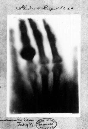
Hand mit Ringen (Hand with Ring): Wilhelm Röntgen's first "medical" X-ray, of his wife's hand, taken on 22 December 1895
X-rays, also known as Röntgen radiation, are short wavelength, high frequency waves. They are a form of electromagnetic radiation. The wavelengths are from around 10-8 to 10-12 meters, with frequencies around 1016 to 1020 hertz.[1]
German physicist Wilhelm Konrad Röntgen discovered X-rays in 1895 while studying electron beams. He found that X-rays could pass through solid objects and cause screens to glow.[1][2] Researchers like Marie Curie, William Coolidge, and Max von Laue expanded his work.[2]
Doses are measured in rems. The SI counterpart is the sievert (Sv).
How X-rays work
X-rays are ionizing radiation, intense enough to break electrons free of their atoms. This can be harmful to the human body, altering genes, chromosomes, and cells.[1] Thus, humans should limit their exposure. (However, modern medical X-rays use very low doses.[3])
Lower-energy X-rays are called "soft X-rays," while higher-energy ones are called "hard X-rays."[2]
Humans can create X-rays with particles. X-rays also emit from stars, supernova remnants, and other space phenomena.[1]
Creating X-rays
There are three main ways to create X-rays:[1]
- Accelerate a charged particle
- Transition an atom between discrete energy levels
- Trigger radioactive decay of atomic nuclei
In X-ray tubes, beams of fast-moving electrons strike metal plates, which deflect and slow the electrons. This causes the electrons to emit "braking radiation," creating X-rays.[1]
Synchrotron particle accelerators are a more powerful way to create X-rays. Charged particles are brought to high speeds, then confined by strong magnets, causing them to emit radiation.[1]
Usage
X-rays have been used to advance scientific understanding. For example, researcher Rosalind Franklin used X-rays to photograph DNA, including her famous Photo 51. This helped reveal how DNA is structured.[2] They can also help scientists study astronomical phenomena like black holes and supernovae.[2]
X-rays can be used to view the structure of crystals.[2]
Healthcare
X-rays are often used in healthcare for medical imaging. They pass through soft tissue while being absorbed by dense body parts like bone and tumors. This makes it easy to diagnose things like dental cavities, fractured bones, and cancer.[1][3] X-ray radiation treatments are sometimes used to damage tumors.[1] Medical X-rays use very low doses of radiation which are not harmful to children and adults.[3]
X-rays can also help with examining objects. For example, airlines scan luggage with X-rays, and manufacturers use X-rays to scan for flaws in castings.[1]